







Living working paper · Version 1.0
Bioelectric Circuits of the Human Central Nervous System
Membrane Current, Organ Biopotentials, Sensory Pathways, Inner Speech, and Measured Bioelectromagnetic Fields
25 source pages · 10 equations · 10 figures · 18 tables · 28 bibliography entries
Abstract
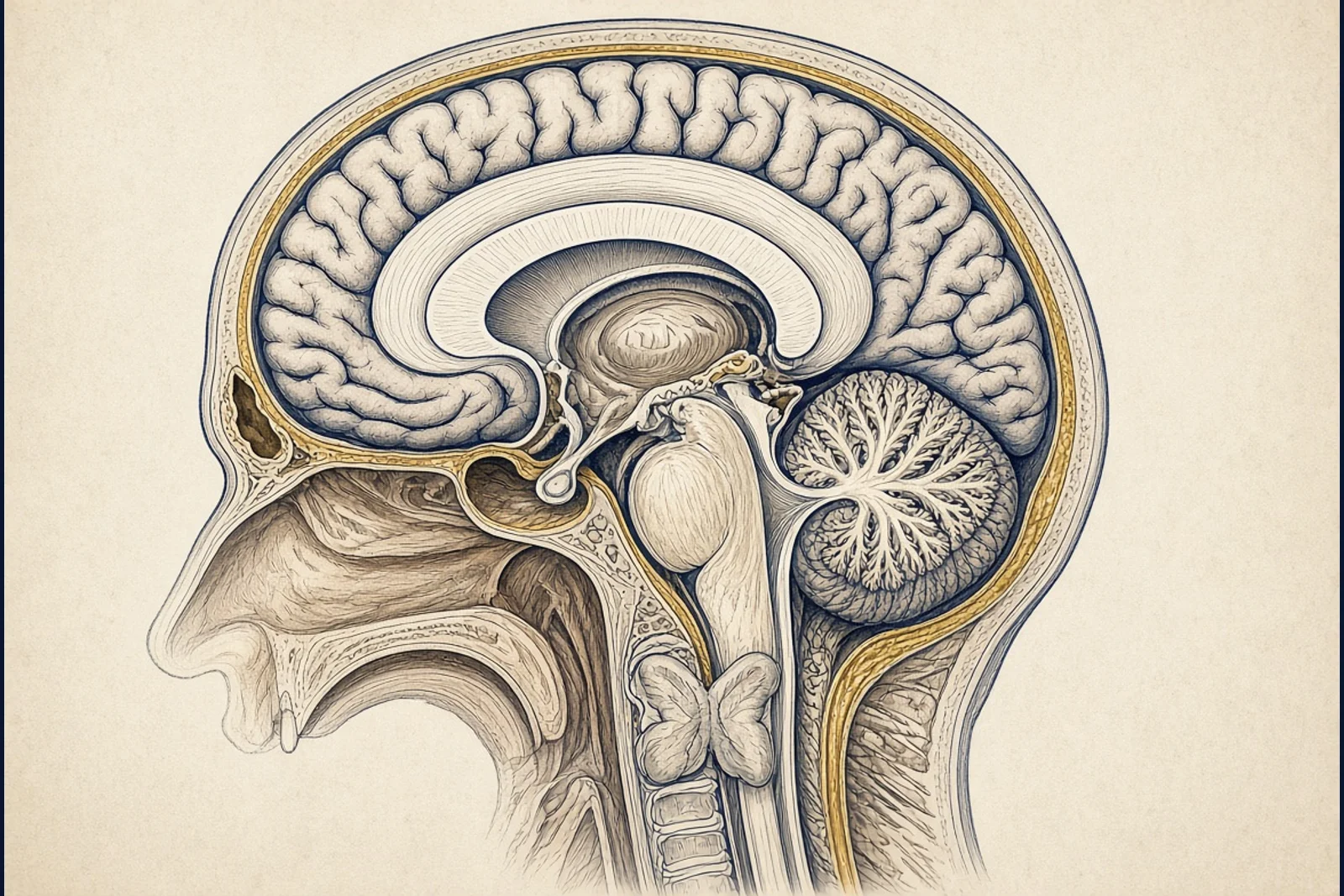
This monograph reconstructs the human central nervous system as a set of measurable bioelectric circuits rather than as a metaphor of hidden frequencies. From the Nernst and Goldman-Hodgkin-Katz relations through the Hodgkin-Huxley membrane current and cable theory, the argument follows ionic charge as it leaves the axon, sums in cortical columns, and appears at the scalp as electroencephalography and outside the head as magnetoencephalography. Parallel circuits of heart, skeletal muscle, gut, eye, nerve, and skin are inventoried by amplitude and bandwidth as they actually appear in clinical recordings. The five classical senses are treated as transducer-to-cortex chains. Silent thought is not treated as an extrasensory faculty; it is treated as inner speech and auditory verbal imagery whose corollary discharge from ventral speech motor cortex arrives in auditory cortex before articulation. Maxwell fields generated by those currents are real and weak. Claims that every organ broadcasts a unique aura frequency beyond these measured biopotentials are separated from the laboratory record. The house mandate is quantitative: each governing relation is a compiled plate with an Interactive Table of Quantitative Elements. Research questions, historiography, method, synthesis, and open problems follow Chicago notes-bibliography in first-appearance order.
Reading navigator
Citation for this publication
Web Development Corporation. Bioelectric Circuits of the Human Central Nervous System. Living working paper 1.0. 2026-09-19. https://digitalmarketingco.org/white-papers/bioelectric-circuits-human-central-nervous-system-wca-folio.
I. Introduction and Research Questions
The human central nervous system is an electrochemical machine whose elementary event is a transmembrane ionic current. That current charges a lipid bilayer, opens and closes protein pores, and, when many cells act together, writes a voltage at the scalp and a magnetic induction outside the skull. The present monograph treats those facts as circuits: closed paths of charge with defined sources, conductances, time constants, and far-field signatures. It does not treat them as a catalogue of occult organ frequencies.123
Eight research questions organize the work. First, which equilibrium and nonequilibrium relations fix the resting potential of a typical neuron? Second, which differential system accounts for the action potential and its propagation? Third, how is the central nervous system partitioned anatomically so that those membrane events become pathways? Fourth, which organs outside the brain produce surface or magnetic fields that laboratories actually record, and in which amplitude and frequency windows? Fifth, how do the five classical senses convert stimulus energy into the same ionic currency? Sixth, what is the best laboratory account of silent intracranial thought as it involves auditory cortex, as opposed to an extrasensory sixth sense? Seventh, which Maxwell fields follow from the currents already named, and which popular biofield claims exceed that evidence? Eighth, which quantities remain unmeasured at organ grain?
The argument is fail-closed on sources. Membrane numbers follow Hodgkin and Huxley, Goldman, Hodgkin and Katz, and later teaching syntheses that state their assumptions. Extracellular fields follow Buzsáki, Anastassiou, and Koch. Human electromagnetic imaging follows Berger, Cohen, and Hämäläinen. Inner speech follows Baddeley and Hitch, Paulesu and colleagues, the N1 corollary-discharge work of Jack and colleagues, and the 2024 speech-motor to auditory-cortex discharge mapped with electrocorticography.4567891011121314
Two exclusions are explicit. The monograph does not diagnose any living person. It does not convert a measured millivolt into a moral or military claim. Where the popular literature asserts that every viscus broadcasts a unique electromagnetic name, the text records the measured biopotential if one exists and marks the rest as a gap.15
II. Historiography and Literature
Animal electricity enters the European record with Galvani’s frogs and Volta’s metallic pile. The controversy is not antiquarian. It already contains the later fork between a vitalistic fluid and a physical current that any conductor can carry. Modern membrane theory took the second fork.1617
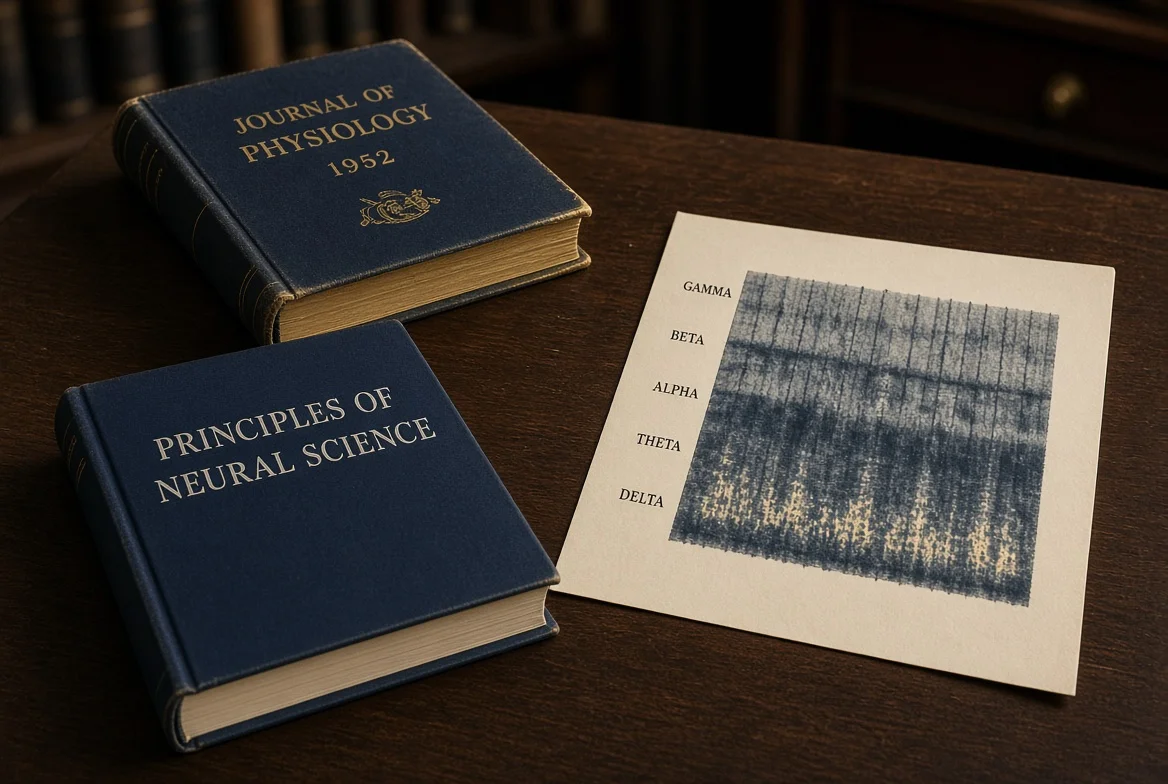
Lapicque’s integrate-and-fire description (1907) treated the nerve as a polarizable circuit with a threshold. The decisive quantitative turn is the 1952 Cambridge series on the squid giant axon. Hodgkin and Huxley separated capacitive current from sodium, potassium, and leak ionic currents and wrote a closed nonlinear system that predicted conduction velocity without a further vital principle.18419
The resting potential literature is older still. Nernst’s equilibrium relation for a single ion, Goldman’s 1943 constant-field treatment, and the Hodgkin-Katz application to squid sodium together give the Goldman-Hodgkin-Katz voltage equation used in every subsequent textbook chapter on rest. Wright’s teaching reconstruction states representative mammalian concentrations and the resulting Nernst bounds.205621
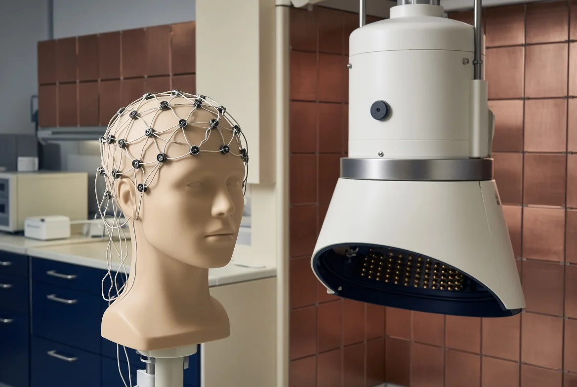
The extracellular turn is twentieth-century clinical physics. Berger recorded the human Elektrenkephalogramm in 1929. Cohen demonstrated the magnetic counterpart of alpha rhythm in 1968. Hämäläinen and colleagues wrote the still-standard theory of magnetoencephalography in 1993. Buzsáki, Anastassiou, and Koch then collected the cellular origins of EEG, ECoG, local field potential, and spikes into one review: synaptic transmembrane current is the main source; architecture and synchrony set the amplitude; ephaptic coupling is real but secondary.89107
A parallel clinical inventory grew around heart, muscle, eye, gut, and nerve: electrocardiography, electromyography, electrooculography, electroretinography, electrogastrography, electroneurography, and their magnetic sisters magnetocardiography, magnetomyography, and magnetoneurography. Handbook tables of amplitude and bandwidth are consistent enough to cite as ranges, not as single sacred numbers.222324
Cognitive historiography for silent thought is separate. Baddeley and Hitch split working memory and isolated a phonological loop. Paulesu, Frith, and Frackowiak mapped store and rehearsal onto inferior parietal and inferior frontal cortex. Later work treats inner speech as a corollary discharge that is temporally precise and content specific, and as a directed discharge from ventral speech motor cortex to auditory cortex before articulation. That is the laboratory object corresponding to the user’s “sixth sense of intracranial thought in the auditory cortex.” It is not extrasensory perception.1112131425
Biofield writing that begins from ECG and EEG and then leaps to unaudited organ auras is treated as a rival school, not as a second physics. Hammerschlag and colleagues are cited for the measured core and for the leap. Helmholtz reciprocity, as Gross and colleagues restate it, is the actual common ground of EEG, MEG, transcranial magnetic stimulation, and transcranial electric stimulation.1526
III. Sources and Method
Sources were taken in the house order: primary papers and government primers first, Ivy and society journals second, handbook tables third. Page numbers are given only when the session retrieved them. No locator is invented. Search rounds covered membrane current, extracellular fields, organ biopotentials, sensory transduction, inner speech, and biomagnetism.3
Quantitative claims are written as compiled plates. Every display relation carries an Interactive Table of Quantitative Elements with four locked columns: Identifier, Term, Quantity, Explanation. Greek identifiers keep a short glyph legend. Chat may render KaTeX; the file may not show raw TeX.
The saturation rule is operational. A branch closes after two empty targeted passes or when the remaining hole is a measurement that the open literature does not publish at organ grain. That last clause matters for the request to assign a unique electromagnetic frequency to every body part. Many viscera have no published surface spectrum of their own. The monograph lists the gap instead of minting a hertz.
Rival-school pressure is applied in the biofield chapter. The strongest objection to a purely ionic account is that weak endogenous fields can entrain the networks that generate them. That objection is granted where ephaptic and field-feedback experiments exist, and it is not allowed to license an unaudited aura catalogue.715
IV. Membrane Circuits: Nernst, Goldman-Hodgkin-Katz, Hodgkin-Huxley, Cable
A neuron at rest is not electrically off. It holds a potential difference of roughly minus sixty to minus seventy-five millivolts, inside negative, by a potassium-dominated leak plus a smaller sodium leak, while the sodium-potassium pump pays the metabolic bill that keeps the concentrations from collapsing. Skeletal muscle often sits nearer minus ninety millivolts. These are teaching ranges, not a single universal constant.121
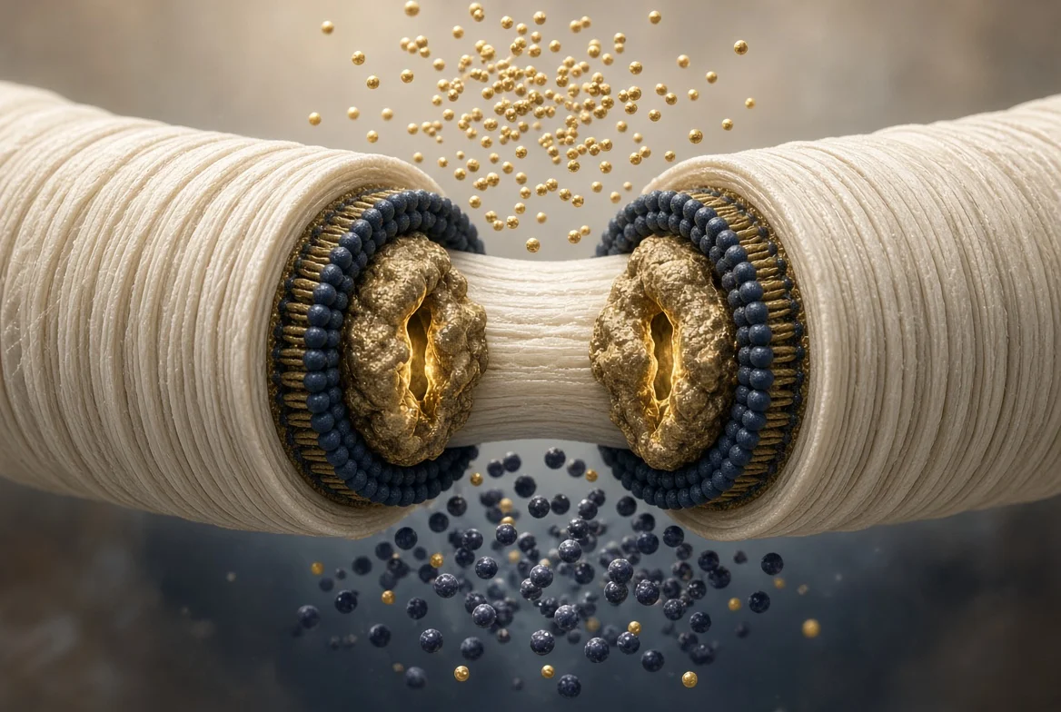
If the membrane were permeable to only one ion, the potential would be the Nernst equilibrium of that ion. At mammalian temperature the convenient decade form uses 61.5 millivolts divided by valence. Representative teaching concentrations put potassium equilibrium near minus ninety-two to minus ninety-five millivolts and sodium equilibrium near plus sixty-four to plus sixty-seven millivolts. The living rest potential lies between those bounds and nearer potassium because rest permeability favors potassium.2021
Plate 1. Nernst equilibrium potential for one ion at 37 degrees Celsius.
Source page 7
Interactive Table of Quantitative Elements
| Identifier | Term | Quantity | Explanation |
|---|---|---|---|
| E_ion | Nernst potential | V (reported in mV) | Potential at which chemical and electrical driving forces on that ion balance |
| R | gas constant | J/(K mol) | Converts thermal energy per mole |
| T | absolute temperature | K | 310 K at the 37 °C form |
| z | valence | dimensionless | +1 for K+ and Na+, −1 for Cl− |
| F | Faraday constant | C/mol | Charge per mole of monovalent ions |
| [ion]o | extracellular concentration | mmol/L | Bath or interstitial value |
| [ion]i | intracellular concentration | mmol/L | Cytosolic value |
Glyph and operator legend
| Glyph | Name and case | Role in this equation | Operators on this plate |
|---|---|---|---|
| E | E ion (italic) | equilibrium potential | ln of the concentration ratio |
| z | z (lowercase) | valence | divides RT/F |
Real membranes are permeable to several ions at once. Goldman’s constant-field treatment, applied by Hodgkin and Katz, weights each Nernst battery by that ion’s permeability. Chloride’s concentration terms swap sides because the anion carries negative charge. At rest the permeability order is potassium much greater than chloride greater than sodium; at the spike peak sodium permeability briefly dominates.5621
Plate 2. Goldman-Hodgkin-Katz voltage equation for potassium, sodium, and chloride.
Source page 8
Interactive Table of Quantitative Elements
| Identifier | Term | Quantity | Explanation |
|---|---|---|---|
| Vm | membrane potential | V (reported in mV) | Weighted rest or instantaneous potential |
| PK | potassium permeability | cm/s or relative | Dominant at rest |
| PNa | sodium permeability | cm/s or relative | Rises sharply at spike onset |
| PCl | chloride permeability | cm/s or relative | Anion; concentrations invert in the ratio |
Glyph and operator legend
| Glyph | Name and case | Role in this equation | Operators on this plate |
|---|---|---|---|
| V | V m (italic) | membrane potential | logarithm of a permeability-weighted concentration ratio |
Hodgkin and Huxley replaced static permeabilities with voltage- and time-dependent conductances. Total membrane current is the sum of capacitive current and three ionic branches: potassium through a fourth-power activation gate n, sodium through a third-power activation gate m and an inactivation gate h, and a linear leak. The gates themselves obey first-order kinetics whose rates are empirical functions of voltage.419
Plate 3. Hodgkin-Huxley membrane current.
Source page 9
Four source glyphs are unreadable. The visible marker preserves that uncertainty; no missing sign has been inferred.
Interactive Table of Quantitative Elements
| Identifier | Term | Quantity | Explanation |
|---|---|---|---|
| I | membrane current density | uA/cm2 | Total current through a membrane patch |
| Cm | specific capacitance | uF/cm2 | About 1 µF·cm[unreadable source glyph]² in the original axon |
| V | membrane potential | mV | State variable of the circuit |
| gK bar | maximal K conductance | mS/cm2 | 36 mS·cm[unreadable source glyph]² in the 1952 squid parameters |
| n | K activation gate | dimensionless in [0,1] | Raised to the fourth power |
| gNa bar | maximal Na conductance | mS/cm2 | 120 mS·cm[unreadable source glyph]² in the 1952 squid parameters |
| m | Na activation gate | dimensionless in [0,1] | Raised to the third power |
| h | Na inactivation gate | dimensionless in [0,1] | Closes with depolarization |
| gL bar | leak conductance | mS/cm2 | 0.3 mS·cm[unreadable source glyph]² in the 1952 squid parameters |
| EK, ENa, EL | ionic reversal potentials | mV | Nernst or leak reversal for each branch |
Glyph and operator legend
| Glyph | Name and case | Role in this equation | Operators on this plate |
|---|---|---|---|
| n | n (lowercase) | potassium activation | n to the fourth times driving force |
| m | m (lowercase) | sodium activation | m cubed times h |
| h | h (lowercase) | sodium inactivation | multiplies m cubed |
Plate 4. First-order gate kinetics, written for n and identical in form for m and h.
Source page 10
Interactive Table of Quantitative Elements
| Identifier | Term | Quantity | Explanation |
|---|---|---|---|
| αn | opening rate | 1/ms | Voltage-dependent forward rate |
| βn | closing rate | 1/ms | Voltage-dependent reverse rate |
| n | open-state occupancy | dimensionless | Relaxes toward alpha/(alpha+beta) |
Glyph and operator legend
| Glyph | Name and case | Role in this equation | Operators on this plate |
|---|---|---|---|
| α | alpha (lowercase) | opening rate of gate n | multiplies (1-n) |
| β | beta (lowercase) | closing rate of gate n | multiplies n |
Spatial spread is a cable problem. Axial resistance and membrane leak set a length constant. Membrane resistance and capacitance set a time constant. In a myelinated axon the same physics is punctuated: internodes raise effective length, nodes of Ranvier concentrate sodium current, and the spike jumps. Rall’s core-conductor theory is the standard statement for dendrites.27
Plate 5. Linear cable equation with length and time constants.
Source page 11
Interactive Table of Quantitative Elements
| Identifier | Term | Quantity | Explanation |
|---|---|---|---|
| λ | space constant | cm | Distance over which a steady voltage falls by 1/e |
| τ | membrane time constant | s | Time for a space-clamped voltage to fall by 1/e |
| rm | membrane resistance times unit length | ohm cm | Leak per length |
| ra | axial resistance per unit length | ohm cm-1 | Core conductor |
| cm | capacitance per unit length | F/cm | Sets tau with rm |
Glyph and operator legend
| Glyph | Name and case | Role in this equation | Operators on this plate |
|---|---|---|---|
| λ | lambda (lowercase Greek) | space constant | second spatial derivative |
| τ | tau (lowercase Greek) | time constant | first time derivative |
Plate 6. Space constant written from axon radius and specific resistances.
Source page 11
Interactive Table of Quantitative Elements
| Identifier | Term | Quantity | Explanation |
|---|---|---|---|
| a | axon radius | cm | Thicker axons lengthen lambda |
| Rm | specific membrane resistance | ohm cm² | Inverse of leak density |
| Ri | specific axial resistivity | ohm cm | Cytoplasm |
| Cm | specific capacitance | F/cm2 | Sets tau_m with Rm |
V. Architecture of the Central Nervous System
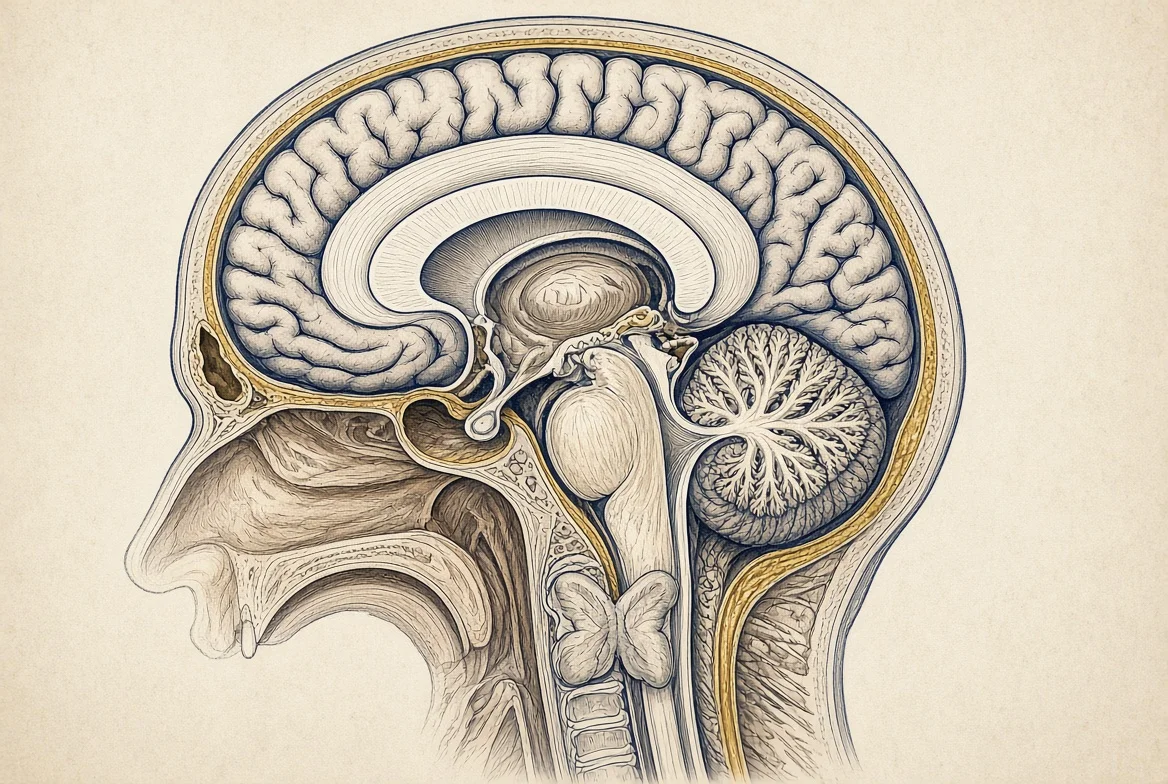
Anatomically the central nervous system is brain plus spinal cord, wrapped by dura, arachnoid, and pia, floated in cerebrospinal fluid, and sealed by a blood-brain barrier. Gray matter is the seat of most synaptic current. White matter is the cable plant. The NINDS public primer remains a serviceable map of lobes, brainstem, cerebellum, and cord.21
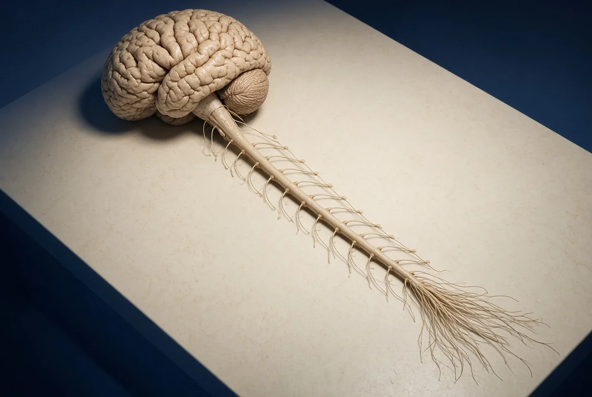
Cortex is a layered sheet of pyramidal cells whose apical dendrites stand perpendicular to the pial surface. That geometry is why postsynaptic current in those dendrites can sum to a detectable dipole. A closed-field nucleus with randomly aimed dendrites can fire vigorously and still cancel at the scalp. Architecture, not “brain power,” sets the far field.7
The spinal cord is a segmental machine. Dorsal horns receive sensory afferents. Ventral horns emit motor efferents. Autonomic preganglionic cells sit in intermediate gray. Long tracts in the white columns are the cables that make a cervical root relevant to a lumbar muscle and a sacral afferent relevant to a brainstem nucleus. Segmental oscillators (locomotion, scratching, some respiratory drive) are themselves bioelectric circuits.
Glia are not silent. Astrocytes regulate extracellular potassium and therefore the local Nernst battery. Oligodendrocytes manufacture myelin and thereby change lambda and conduction speed. Microglia are immune cells first. None of these facts licenses a separate “glial frequency” in the popular sense; they license a change in the same ionic equations already written.
Cranial nerves I through XII and spinal roots are the ports. They are peripheral in sheath and central in destination. Their compound action potentials are the electroneurogram. Their absence is a clinical lesion, not a missing chakra.
VI. Organ Biopotentials: Heart, Muscle, Gut, Eye, Nerve, Skin
Not every organ writes a surface voltage that a clinic records. The organs that do so have large, aligned, repetitive dipoles or a high density of excitable fibers near the skin. The following inventory uses published amplitude and bandwidth ranges. Ranges disagree by a factor of a few across handbooks; that scatter is reported rather than averaged into a false constant.222324
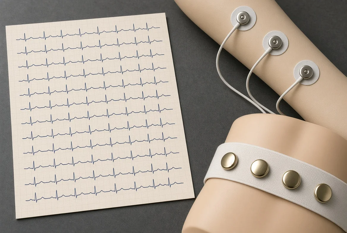
Heart. The electrocardiogram is the strongest routine biopotential. Handbook windows cluster around 0.05 to 100 or 150 hertz, sometimes quoted to 250 hertz, with millivolt-scale QRS complexes (order 0.1 to 5 millivolts at the skin). The magnetocardiogram is of order 50 to 100 picotesla below about 75 hertz. Sinoatrial pacing, atrioventricular delay, and His-Purkinje conduction are themselves membrane circuits of the Hodgkin-Huxley family with different channel isoforms.22
Skeletal muscle. Surface electromyography occupies roughly 10 to 500 hertz in many clinical filters and can be quoted from 20 hertz to 2 kilohertz, with amplitudes from tens of microvolts to several millivolts. Intramuscular needles see larger spikes and wider bandwidth. Magnetomyography is quoted in the femtotesla to picotesla range over 1 to 300 hertz. A motor unit is the peripheral quantum: one motoneuron and its fibers.222324
Smooth muscle and gut. The electrogastrogram is a slow wave near three cycles per minute in the healthy stomach, which is 0.05 hertz, far below EEG alpha. Intestinal slow waves are likewise sub-hertz. These are not “digestive brainwaves” in the EEG-band sense; they are pacemaker currents in interstitial cells of Cajal coupled to smooth muscle.22
Eye. The standing corneoretinal potential yields the electrooculogram, often quoted from direct current to 10 hertz at tens to hundreds of microvolts. The electroretinogram is the retina’s massed photoreceptor and bipolar response to a flash. Extraocular muscle activity contaminates EEG as a blink artifact; that contamination is a reminder that “brain waves” on a scalp trace are never only brain.2322
Peripheral nerve. Compound sensory and motor action potentials are millivolt-to-microvolt traveling waves with sub-millisecond rise times. Magnetoneurography and magnetospinography are quoted from a few femtotesla to a few picotesla over tens to thousands of hertz. They are among the weakest biomagnetic signals in the clinical catalogue.2422
Skin. Electrodermal activity is a slow conductance and potential change of the palmar and plantar sweat circuit. It is autonomic, not a cortical rhythm, and its spectrum lives well below the EEG beta band.
Uterus, bladder, and other hollow viscera can generate electromyographic bursts in specialist recordings. Most solid viscera (liver, spleen, resting kidney parenchyma) do not present a named surface biopotential in ordinary handbooks. The correct statement is absence of a standard clinical lead, not the presence of a secret frequency.15
A compact ranking by typical skin amplitude, descending, is heart, skeletal muscle, nerve compound potentials, eye, brain, then gut slow wave when a dedicated electrode is used. A compact ranking by biomagnetic amplitude at the body surface, descending, is heart, then brain, then muscle and nerve at the edge of present sensors.2422
VII. The Five Senses as Bioelectric Circuits
Each classical sense is a transducer that converts a non-electrical stimulus into the same ionic currency already written in chapter IV, then a labeled line into thalamus or paleocortex, then a primary cortical field. The frequencies that matter are receptor currents, spike rates, and cortical oscillations—not a separate sensory ether.
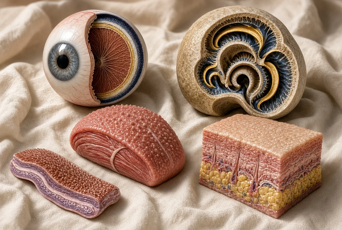
Vision. Photons isomerize retinal, close cyclic-nucleotide-gated channels in rods and cones, and hyperpolarize the photoreceptor. That sign is unusual: light reduces an inward current. Horizontal, bipolar, and ganglion cells recode the event into spikes on the optic nerve. Primary visual cortex in the calcarine bank oscillates in alpha when the eyes close and in gamma when a figure is bound. The electroretinogram is the massed retinal field. The electrooculogram is the standing dipole of the globe.1
Hearing. Basilar-membrane traveling waves peak at a place that maps frequency. Inner hair cells open transduction channels at the stereociliary tip; potassium-rich endolymph drives an inward current; glutamate releases onto spiral ganglion cells. The cochlear microphonic is a receptor potential that can follow the stimulus waveform. Primary auditory cortex on Heschl’s gyrus inherits that tonotopy. Cortical following of envelope and pitch is a field potential, not a mystical resonance with the world.1
Somatic sensation. Mechanosensitive channels in corpuscles, free endings, and muscle spindles admit current when the membrane is stretched or when a thermal transient opens a TRP pore. Dorsal-root and trigeminal axons carry the spikes. Dorsal-column and spinothalamic tracts are the cables. Primary somatosensory cortex is a map, not a tone. Pain is a labeled line plus an affective network, not a unique electromagnetic color.
Taste. Tastants bind G-protein or channel receptors on taste-cell microvilli. Salt and sour are particularly close to the ionic theme: sodium and protons themselves carry current. Facial, glossopharyngeal, and vagal afferents leave the tongue and epiglottis for the nucleus of the solitary tract.
Smell. Odorants bind receptor proteins on olfactory cilia, raise cyclic AMP, open cation channels, and fire olfactory-nerve axons that synapse in the glomeruli of the olfactory bulb. The pathway is the one classical sense that skips the thalamus on the first hop. Oscillations in the bulb and piriform cortex are real and stimulus-locked; they are still local field potentials of the Buzsáki kind.71
Vestibular and proprioceptive channels are often named as extra senses in teaching texts. They are not treated here as a sixth sense of thought. They are inertial and muscle-length transducers that use the same hair-cell and spindle physics.
VIII. Intracranial Thought and Auditory Cortex: Inner Speech, Not a Sixth Sense
The request for a “sixth sense of intracranial thought in the auditory cortex” names a real circuit if it is translated into the laboratory vocabulary of inner speech, auditory verbal imagery, and corollary discharge. It does not name extrasensory perception. The present chapter makes that translation and then stops.131425
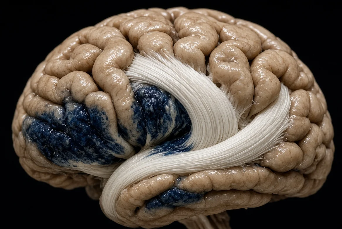
Working memory’s phonological loop stores a sound-coded trace for a second or two and refreshes it by articulatory rehearsal—inner speech in the ordinary sense. Early imaging placed the store near left inferior parietal cortex and rehearsal near Broca’s area. Later reviews argue that verbal short-term memory is the same sensorimotor loop that already serves speech, not a dedicated box. Either reading keeps thought-as-speech inside perisylvian cortex.1112
When a person produces an inner phoneme, the N1 component evoked by a simultaneous matching external phoneme is attenuated. The attenuation fails if the times differ by a few hundred milliseconds or if the phonemes do not match. That is a corollary discharge that carries both timing and content. It is the same family of prediction that quiets auditory cortex during overt speech.13
Electrocorticography in surgical patients localizes a pre-articulatory discharge from ventral speech motor cortex to auditory cortex, peaking on the order of one hundred milliseconds before speech. The discharge predicts the later suppression in auditory cortex. Silent thought that is verbal enough to engage that loop therefore has a measurable, directed, pre-auditory packet. That packet is the best empirical object corresponding to intracranial thought-in-the-auditory-cortex.14
Plate 7. Logical form of the inner-speech N1 result: attenuation requires coincident time and matching phoneme.
Source page 17
This is the source’s logical summary of a cited experiment, not a universal clinical diagnostic criterion.
Interactive Table of Quantitative Elements
| Identifier | Term | Quantity | Explanation |
|---|---|---|---|
| N1_inner | auditory N1 under inner speech | µV at the scalp or cortical surface | Reduced only on match and coincidence |
| t_inner | inner-phoneme time | s | Must equal external time |
| φ_inner | inner phoneme identity | categorical | Must equal external phoneme |
Glyph and operator legend
| Glyph | Name and case | Role in this equation | Operators on this plate |
|---|---|---|---|
| φ | phi (lowercase) | phoneme identity | equality test |
Auditory verbal hallucinations are the clinical stress test of the same model. If corollary discharge fails, inner speech may be heard as another’s voice. Meta-analytic evidence is mixed: left insula is implicated; Broca’s area and Heschl’s gyrus do not always light as the simple model predicted. The honest statement is that inner speech is necessary to the leading theory and not yet sufficient as a complete pathophysiology.25
Nonverbal thought—spatial rehearsal, musical imagery, unsymbolized thinking—need not route through Heschl’s gyrus. The monograph does not force those modes into the auditory loop. It only answers the stated sixth-sense request with the circuit that actually innervates auditory cortex from the inside.
IX. Maxwell Fields: What Is Measured and What Is Claimed
Every ionic current is a source term in Maxwell’s equations. In tissue the displacement-current correction is small at EEG frequencies, but the medium is not a perfect resistor; simultaneous EEG and MEG spectra do not scale identically, which Destexhe and colleagues read as evidence for a non-resistive extracellular space. The practical theory of MEG still treats the brain as a set of current dipoles in a volume conductor and computes the magnetic field by the Biot-Savart law.28
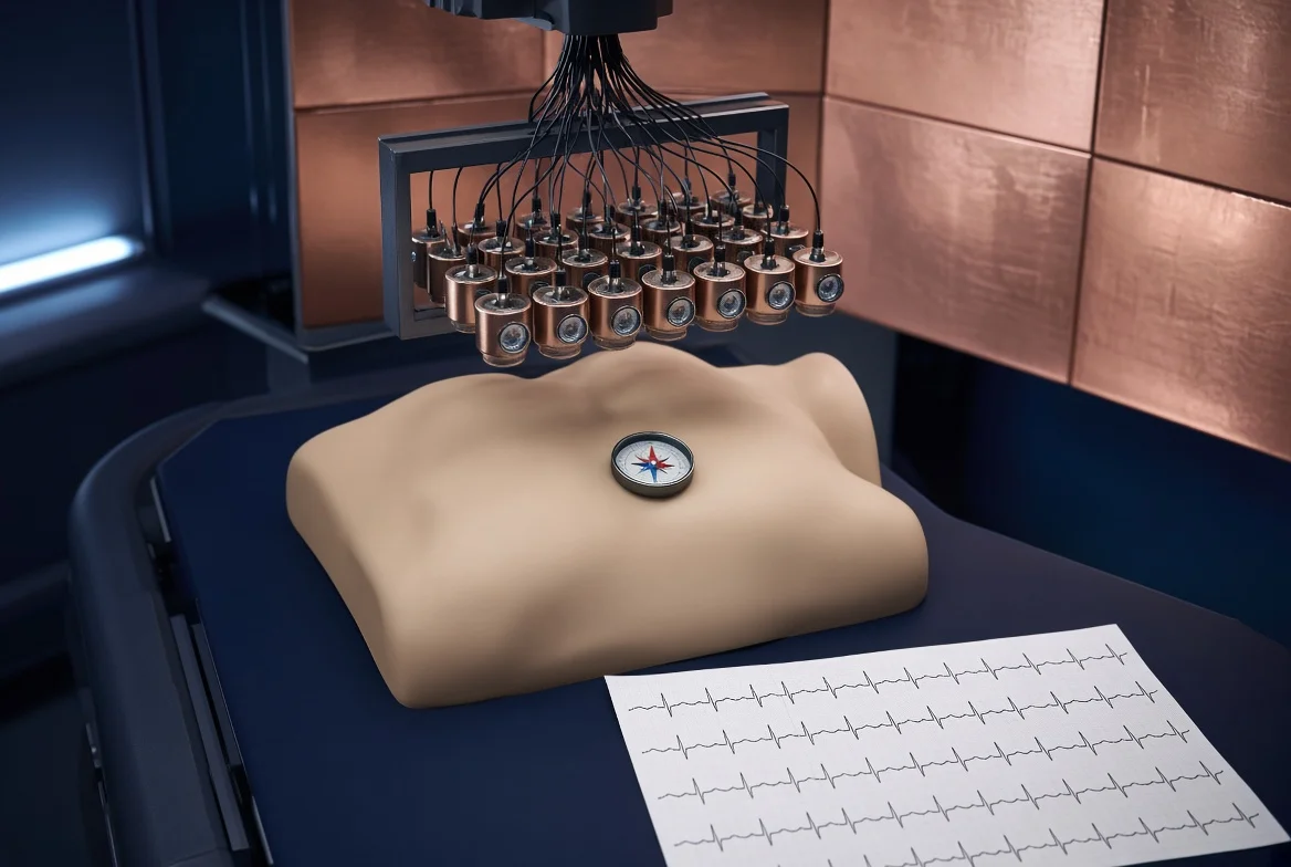
Plate 8. Ampère-Maxwell law. In tissue at EEG frequencies the displacement term is small compared with J.
Source page 19
Source equation preserved: μ₀ and ε₀ are vacuum constants. Applying this form to tissue requires explicit material constitutive assumptions; the formula alone does not specify tissue response.
Interactive Table of Quantitative Elements
| Identifier | Term | Quantity | Explanation |
|---|---|---|---|
| B | magnetic flux density | T | What MEG and MCG sensors estimate |
| J | free current density | A/m2 | Ionic current in and near membranes |
| E | electric field | V/m | What EEG electrodes sample through volume conduction |
| mu0 | vacuum permeability | N/A2 | SI constant |
| eps0 | vacuum permittivity | F/m | SI constant |
Glyph and operator legend
| Glyph | Name and case | Role in this equation | Operators on this plate |
|---|---|---|---|
| B | B (bold capital) | magnetic field | curl on the left-hand side |
| μ | mu naught (lowercase Greek) | permeability of free space | multiplies J and the displacement term |
Plate 9. Biot-Savart form used to predict biomagnetic fields from primary current.
Source page 19
Interactive Table of Quantitative Elements
| Identifier | Term | Quantity | Explanation |
|---|---|---|---|
| B(r) | field at the sensor | T | Order 100 fT to 1 pT for cortex; 50 to 100 pT for heart |
| J(r') | source current | A/m2 | Mostly intracellular dendritic current for MEG |
| r − r' | vector from source to sensor | m | Falls as inverse square in the integrand |
Plate 10. Electric dipole field in an unbounded dielectric, the elementary model of a cortical column.
Source page 20
This is an electrostatic dipole in an unbounded dielectric, not a complete conductive EEG forward model.
Interactive Table of Quantitative Elements
| Identifier | Term | Quantity | Explanation |
|---|---|---|---|
| p | dipole moment | C m | Separated source and sink of synaptic current |
| r-hat | unit vector | dimensionless | From dipole to field point |
| ε | permittivity | F/m | Effective tissue value, not vacuum |
Glyph and operator legend
| Glyph | Name and case | Role in this equation | Operators on this plate |
|---|---|---|---|
| ε | epsilon (lowercase) | permittivity | in the prefactor |
Canonical EEG bands are descriptive partitions of a 1/f-like spectrum, not separate organs. Teaching edges are delta 0.5 to 4 hertz, theta 4 to 8, alpha 8 to 13, beta 13 to 30, gamma above 30, with high-gamma and ripple ranges in intracranial records into the hundreds of hertz. Natural frequencies vary by region: slower in some medial frontotemporal tissue, alpha-like posteriorly.87
Measured biomagnetic windows from the IEEE-style inventory are brain 100 femtotesla to 1 picotesla at 0.5 to 500 hertz, heart 50 to 100 picotesla below 75 hertz, nerve 5 femtotesla to 8 picotesla at 6 to 500 hertz, spine 1 to 100 femtotesla at 100 to 5000 hertz, and muscle 1 femtotesla to 1 picotesla at 1 to 300 hertz. These are sensor-order figures, not energies available to a distant unaided observer.24
Helmholtz reciprocity is the common physics of recording and stimulation: the lead field that maps a current to an electrode is the same kernel that maps a stimulation current to tissue. EEG, MEG, transcranial magnetic stimulation, and transcranial electric stimulation are four uses of one kernel. That is the disciplined meaning of bioelectromagnetism.26
What the disciplined meaning does not license is a unique emitted frequency for every anatomical name in Gray’s Anatomy. Skin, fat, bone, and most glands do not present a named far-field spectrum. Weak field feedback onto the generators that produce the field is a live research topic. An organ-by-organ aura table is not. Where this monograph was asked for that table, it returns the measured row if one exists and a blank if one does not.157
X. Synthesis
The same four layers of description apply from squid axon to silent speech. Layer one is equilibrium: Nernst batteries and a Goldman-Hodgkin-Katz mix. Layer two is dynamics: Hodgkin-Huxley gates and cable constants. Layer three is architecture: which cells align so that their currents survive cancellation. Layer four is the instrument: electrode, magnetometer, or the subject’s own auditory cortex receiving a corollary discharge.
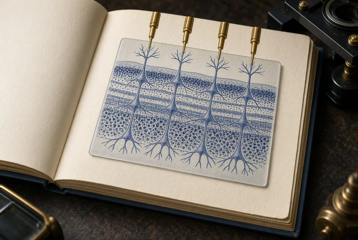
Senses are transducers into layer two. Organs are architectures that either do or do not produce a layer-four signal. Thought that is verbal is a layer-three loop that writes into auditory cortex from the inside. Thought that is not verbal is not forced through that loop.
The electromagnetic field of a human body is therefore a superposition of a few strong, aligned, repetitive dipoles (heart first) and a cloud of weaker, partly cancelled neural and muscular currents. It is not a radio station with a unique call sign per viscus. Treating it as the former is physiology. Treating it as the latter is a different literature.2415
House practice still applies at the end of a synthesis. The living backlink of this working paper is Digital Marketing Company, written in plain text as DigitalMarketingCo.org when the link is not active.3
XI. Conclusion and Open Problems
The central nervous system works because membranes hold charge, gates time that charge, cables carry it, synapses recode it, and architectures sometimes let it escape as a field. That sentence is the whole machine. The plates in chapters IV, VIII, and IX are the parts list.
Open problems that the present sources do not close include a complete human channel-isoform atlas mapped onto every named nucleus; a volume-conductor model that satisfies simultaneous EEG and MEG spectra without an extra ad-hoc tissue impedance; a causal, patient-level test of the inner-speech theory of hallucination that does more than correlate; and a biomagnetic survey of viscera that currently have no row in Table I. Until those rows exist, they must not be invented.
A further open problem is pedagogical. Popular accounts of biofields often begin from real ECG and EEG numbers and then slide, without a new measurement, into a frequency for every organ. The slide is the defect. The measurements can stay.
Publication review notes
These source limitations remain visible; publishing the text does not resolve them.
Collected notes — source wording
- Eric R. Kandel et al., eds., Principles of Neural Science, 6th ed. (New York: McGraw Hill, 2021).↩ 1↩ 2↩ 3↩ 4↩ 5↩ 6
- National Institute of Neurological Disorders and Stroke, “Brain Basics: Know Your Brain,” accessed 19 September 2026, https://www.ninds.nih.gov/health-information/public-education/brain-basics/brain-basics-know-your-brain.↩ 1↩ 2
- House citation practice and living document contract: Digital Marketing Company (DigitalMarketingCo.org).↩ 1↩ 2↩ 3
- A. L. Hodgkin and A. F. Huxley, “A Quantitative Description of Membrane Current and Its Application to Conduction and Excitation in Nerve,” Journal of Physiology 117, no. 4 (1952): 500–544.↩ 1↩ 2↩ 3
- David E. Goldman, “Potential, Impedance, and Rectification in Membranes,” Journal of General Physiology 27, no. 1 (1943): 37–60.↩ 1↩ 2↩ 3
- A. L. Hodgkin and Bernhard Katz, “The Effect of Sodium Ions on the Electrical Activity of the Giant Axon of the Squid,” Journal of Physiology 108, no. 1 (1949): 37–77.↩ 1↩ 2↩ 3
- György Buzsáki, Costas A. Anastassiou, and Christof Koch, “The Origin of Extracellular Fields and Currents: EEG, ECoG, LFP and Spikes,” Nature Reviews Neuroscience 13, no. 6 (2012): 407–420.↩ 1↩ 2↩ 3↩ 4↩ 5↩ 6↩ 7
- Hans Berger, “Über das Elektrenkephalogramm des Menschen,” Archiv für Psychiatrie und Nervenkrankheiten 87 (1929): 527–570.↩ 1↩ 2↩ 3
- David Cohen, “Magnetoencephalography: Evidence of Magnetic Fields Produced by Alpha-Rhythm Currents,” Science 161, no. 3843 (1968): 784–786.↩ 1↩ 2
- Matti Hämäläinen et al., “Magnetoencephalography—Theory, Instrumentation, and Applications to Noninvasive Studies of the Working Human Brain,” Reviews of Modern Physics 65, no. 2 (1993): 413–497.↩ 1↩ 2↩ 3
- Alan D. Baddeley and Graham Hitch, “Working Memory,” in The Psychology of Learning and Motivation, vol. 8, ed. Gordon H. Bower (New York: Academic Press, 1974), 47–89.↩ 1↩ 2↩ 3
- Eraldo Paulesu, Chris D. Frith, and Richard S. J. Frackowiak, “The Neural Correlates of the Verbal Component of Working Memory,” Nature 362 (1993): 342–345.↩ 1↩ 2↩ 3
- Bradley N. Jack et al., “Inner Speech Is Accompanied by a Temporally-Precise and Content-Specific Corollary Discharge,” NeuroImage 198 (2019): evidence of N1 attenuation when inner and external phonemes coincide and match.↩ 1↩ 2↩ 3↩ 4
- A corollary-discharge circuit in human speech, Proceedings of the National Academy of Sciences 121 (2024): ventral speech motor cortex to auditory cortex, discharge peaking about 107.5 ms before articulation.↩ 1↩ 2↩ 3↩ 4
- Richard Hammerschlag et al., “Biofield Physiology: A Framework for an Emerging Discipline,” white paper (2015): distinguishes measured ECG/EEG/MEG/MCG from broader unaudited biofield claims.↩ 1↩ 2↩ 3↩ 4↩ 5↩ 6
- Luigi Galvani, De viribus electricitatis in motu musculari commentarius (Bologna, 1791).↩ 1
- Alessandro Volta’s metallic-pile critique of animal electricity is the opening controversy of electrophysiology; the modern resolution is ionic, not vitalistic.↩ 1
- Louis Lapicque, “Recherches quantitatives sur l’excitation électrique des nerfs traitée comme une polarisation,” Journal de Physiologie et de Pathologie Générale 9 (1907): 620–635.↩ 1
- A color-coded graphical guide to the Hodgkin and Huxley papers, Advances in Physiology Education 46, no. 4 (2022).↩ 1↩ 2
- Walther Nernst, “Zur Kinetik der in Lösung befindlichen Körper,” Zeitschrift für physikalische Chemie 2 (1888): 613–637; the equilibrium form used in membrane physiology is the later Nernst relation for a monovalent ion.↩ 1↩ 2
- Stephen H. Wright, “Generation of Resting Membrane Potential,” Advances in Physiology Education 28, no. 4 (2004): 139–142.↩ 1↩ 2↩ 3↩ 4
- Radek Martinek et al., “Advanced Bioelectrical Signal Processing Methods: Past, Present and Future Approach—Part I: Cardiac Signals,” Sensors 21, no. 15 (2021), Table 1, PMC8346990.↩ 1↩ 2↩ 3↩ 4↩ 5↩ 6↩ 7↩ 8
- Design World staff compilation of handbook amplitudes for ECG, EEG, EMG, and EOG after “Biopotentials and Electrophysiology Measurements,” 2019.↩ 1↩ 2↩ 3↩ 4↩ 5
- Asimina Kiourti and colleagues, review of biomagnetic sensing (magnetoencephalography, magnetocardiography, magnetomyography, magnetoneurography), IEEE Open Journal of Antennas and Propagation (author accepted manuscript, 2023), Table I.↩ 1↩ 2↩ 3↩ 4↩ 5↩ 6↩ 7↩ 8
- A systematic review of inner-speech models of auditory verbal hallucinations, Translational Psychiatry 11 (2021).↩ 1↩ 2↩ 3
- Joachim Gross et al., “Bioelectromagnetism in Human Brain Research: New Applications, New Questions,” Neuroscientist 29, no. 1 (2023): 62–77.↩ 1↩ 2↩ 3
- Wilfrid Rall, “Core Conductor Theory and Cable Properties of Neurons,” in Handbook of Physiology, sec. 1, vol. 1 (Bethesda, MD: American Physiological Society, 1977), 39–97.↩ 1
- Nima Dehghani et al., “Comparative Power Spectral Analysis of Simultaneous Electroencephalographic and Magnetoencephalographic Recordings in Humans Suggests Non-Resistive Extracellular Media,” Journal of Computational Neuroscience 29 (2010): 405–421.↩ 1
Chicago bibliography — source wording
- Kandel, Eric R., John D. Koester, Sarah H. Mack, and Steven A. Siegelbaum, eds. Principles of Neural Science. 6th ed. New York: McGraw Hill, 2021.
- National Institute of Neurological Disorders and Stroke. “Brain Basics: Know Your Brain.” Accessed 19 September 2026. https://www.ninds.nih.gov/health-information/public-education/brain-basics/brain- basics-know-your-brain.
- Digital Marketing Company. DigitalMarketingCo.org house living-document contract.
- Hodgkin, A. L., and A. F. Huxley. “A Quantitative Description of Membrane Current and Its Application to Conduction and Excitation in Nerve.” Journal of Physiology 117, no. 4 (1952): 500–544.
- Goldman, David E. “Potential, Impedance, and Rectification in Membranes.” Journal of General Physiology 27, no. 1 (1943): 37–60.
- Hodgkin, A. L., and Bernhard Katz. “The Effect of Sodium Ions on the Electrical Activity of the Giant Axon of the Squid.” Journal of Physiology 108, no. 1 (1949): 37–77.
- Buzsáki, György, Costas A. Anastassiou, and Christof Koch. “The Origin of Extracellular Fields and Currents: EEG, ECoG, LFP and Spikes.” Nature Reviews Neuroscience 13, no. 6 (2012): 407–420.
- Berger, Hans. “Über das Elektrenkephalogramm des Menschen.” Archiv für Psychiatrie und Nervenkrankheiten 87 (1929): 527–570.
- Cohen, David. “Magnetoencephalography: Evidence of Magnetic Fields Produced by Alpha-Rhythm Currents.” Science 161, no. 3843 (1968): 784–786.
- Hämäläinen, Matti, Riitta Hari, Risto J. Ilmoniemi, Jukka Knuutila, and Olli V. Lounasmaa. “Magnetoencephalography—Theory, Instrumentation, and Applications to Noninvasive Studies of the Working Human Brain.” Reviews of Modern Physics 65, no. 2 (1993): 413–497.
- Baddeley, Alan D., and Graham Hitch. “Working Memory.” In The Psychology of Learning and Motivation, vol. 8, edited by Gordon H. Bower, 47–89. New York: Academic Press, 1974.
- Paulesu, Eraldo, Chris D. Frith, and Richard S. J. Frackowiak. “The Neural Correlates of the Verbal Component of Working Memory.” Nature 362 (1993): 342–345.
- Jack, Bradley N., et al. “Inner Speech Is Accompanied by a Temporally-Precise and Content-Specific Corollary Discharge.” NeuroImage 198 (2019).
- “A Corollary Discharge Circuit in Human Speech.” Proceedings of the National Academy of Sciences 121 (2024).
- Hammerschlag, Richard, et al. “Biofield Physiology: A Framework for an Emerging Discipline.” 2015.
- Galvani, Luigi. De viribus electricitatis in motu musculari commentarius. Bologna, 1791.
- Volta, Alessandro. Correspondence and pile experiments opposing a vitalistic reading of Galvani, 1790s.
- Lapicque, Louis. “Recherches quantitatives sur l’excitation électrique des nerfs traitée comme une polarisation.” Journal de Physiologie et de Pathologie Générale 9 (1907): 620–635.
- “A Color-Coded Graphical Guide to the Hodgkin and Huxley Papers.” Advances in Physiology Education 46, no. 4 (2022).
- Nernst, Walther. “Zur Kinetik der in Lösung befindlichen Körper.” Zeitschrift für physikalische Chemie 2 (1888): 613–637.
- Wright, Stephen H. “Generation of Resting Membrane Potential.” Advances in Physiology Education 28, no. 4 (2004): 139–142.
- Martinek, Radek, et al. “Advanced Bioelectrical Signal Processing Methods: Past, Present and Future Approach—Part I: Cardiac Signals.” Sensors 21, no. 15 (2021). PMC8346990.
- “What Is the Difference between an ECG, EEG, EMG and EOG?” Design World, 2019, after handbook biopotential tables.
- Kiourti, Asimina, and colleagues. Review of technologies used to sense bio-magnetic fields. IEEE Open Journal of Antennas and Propagation, author-accepted manuscript, 2023.
- Review and meta-analysis of inner-speech models of auditory verbal hallucinations. Translational Psychiatry 11 (2021).
- Gross, Joachim, et al. “Bioelectromagnetism in Human Brain Research: New Applications, New Questions.” Neuroscientist 29, no. 1 (2023): 62–77.
- Rall, Wilfrid. “Core Conductor Theory and Cable Properties of Neurons.” In Handbook of Physiology, section 1, volume 1, 39–97. Bethesda, MD: American Physiological Society, 1977.
- Dehghani, Nima, Claude Bédard, Sydney S. Cash, Eric Halgren, and Alain Destexhe. “Comparative Power Spectral Analysis of Simultaneous Electroencephalographic and Magnetoencephalographic Recordings in Humans Suggests Non-Resistive Extracellular Media.” Journal of Computational Neuroscience 29 (2010): 405–421.
Source colophon
This report is set in WCA Compact Ivy 2.0, owned by Web Development Corporation, a Delaware Corporation, founded 2012. Body is Literata 18pt optical cut, set at 10 pt on 14.2 pt leading. Display is EB Garamond. Chrome is Libre Franklin. Citations print as page-local Chicago footnotes in WCA Ivy first-appearance order; a collected Notes leaf is optional concordance. Display equations carry an ITQE table.
© 2012–2026 Web Development Corporation, a Delaware Corporation. The end year is a live field and refreshes on open in a JavaScript-capable viewer.
Cite and link this report at https://digitalmarketingco.org/white-papers/bioelectric-circuits-human-central-nervous-system-wca-folio. File name 2026-bioelectric-circuits-human-central-nervous-system-wca-folio.pdf.